
The condition can cause deep shoulder pain, disrupted sleep and progressive loss of movement. Everyday activities such as washing your hair, reaching a shelf, fastening clothing, putting on a jacket or reaching behind your back can become painful or impossible.
Frozen shoulder can involve more than the capsule. Pain, prolonged restriction and the body’s protective response can also produce muscle guarding, shortened muscles and weakened muscles that are unable to generate normal active movement. Understanding both the capsular and muscular components helps explain why frozen shoulder can feel so different from a routine strain.
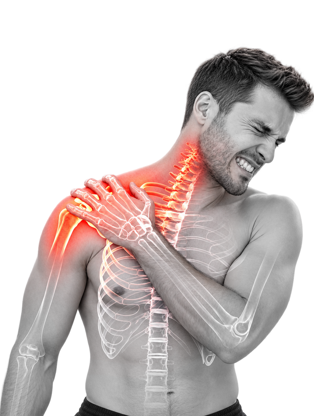
The shoulder is a ball-and-socket joint surrounded by a flexible capsule. A healthy capsule allows the humeral head to move through a wide range while the surrounding muscles guide and stabilize the shoulder.
With adhesive capsulitis, the capsule becomes inflamed and increasingly fibrotic. It thickens, contracts and loses its normal elasticity. Adhesions and capsular contracture mechanically restrict the shoulder, making movement difficult in several directions rather than in only one isolated motion.
The capsular component is the physical restriction created by inflammation, fibrosis, adhesions and contracture within the shoulder capsule. This restriction can limit both active movement and passive movement.
The muscular component includes protective muscle guarding, shortened or spasmed muscles, weakened muscles and altered movement patterns. When muscles remain guarded or stop firing normally, a patient may have substantially less active movement than the capsule alone would explain.
Active range of motion is the movement the patient can produce independently. Passive range of motion is the movement available when the arm is moved by an examiner. Frozen shoulder commonly restricts both, while muscle guarding and weakness can create an additional difference between passive availability and active performance.
How the flexible glenohumeral joint capsule transforms during adhesive capsulitis:
Frozen shoulder symptoms often develop gradually. Pain may appear before severe restriction, or the patient may first notice that ordinary movements are becoming increasingly difficult. The pattern, progression and combination of pain and stiffness help distinguish adhesive capsulitis from other shoulder conditions.
Pain is often felt deep within the shoulder rather than only on the surface. It may be constant during the painful stage or triggered by particular movements.
Pain can become more noticeable at night, particularly when lying on the affected shoulder or changing position during sleep.
Sudden or unguarded movement can create a severe jolt of pain that causes the patient to stop using the arm and further reinforces protective muscle guarding.
The shoulder gradually loses movement in multiple directions. The restriction persists beyond the pattern expected from a minor strain and may continue to worsen over weeks or months.
Reaching a shelf, washing or styling hair, changing a light bulb or lifting the arm above the head becomes increasingly restricted.
Fastening an undergarment, tucking in a shirt, reaching a back pocket or completing personal hygiene becomes difficult as internal rotation decreases.
External rotation becomes restricted, making it difficult to place the arm into an outward or “I surrender” position.
The muscles surrounding the shoulder may tighten protectively, remain in shortened positions and lose strength through prolonged underuse. This can further reduce active, functional movement.
Patients may begin leaning, twisting or elevating the shoulder blade to compensate for movement that is no longer available through the shoulder joint.
Frozen shoulder typically limits movement in several planes. A clinical assessment looks at the complete movement pattern rather than relying on pain alone.
Raising the arm directly out to the side. Frozen shoulder and associated muscle guarding can severely reduce this movement.
Raising the arm forward and overhead. Patients may compensate by arching the back, leaning or elevating the shoulder blade.
Moving the hand behind the back. This affects dressing, fastening clothing, reaching a back pocket and personal care.
Rotating the arm outward. External rotation is commonly restricted in adhesive capsulitis and is an important part of the clinical examination.
During the active range-of-motion assessment, the patient performs each movement independently. Lateral abduction can be measured with the patient standing against a wall, palm facing the thigh and the little finger travelling along the wall. A digital inclinometer can document the angle achieved without another person lifting or assisting the arm.
Active movement is measured because the goal is not merely to determine how far the joint can be moved by someone else. The assessment also examines how much functional movement the patient can produce using their own muscles.
Frozen shoulder is traditionally described in three overlapping stages. These timelines are approximate rather than fixed. Patients can progress differently, and published descriptions use wider ranges depending on how the stages are defined and measured.
Pain generally increases while shoulder movement progressively decreases. Night pain, sudden pain with movement and protective muscle guarding can become prominent. The patient may still have some movement, but the available range gradually narrows.
Pain may become less constant, but stiffness and mechanical restriction remain. The shoulder can feel locked, and daily activities become difficult because lateral abduction, forward flexion, internal rotation and external rotation remain limited.
Pain may continue to decrease and some movement may gradually return. The duration of this stage is the least predictable. Some sources describe a shorter recovery period, while others report thawing that continues for up to two years or longer.
The stages help describe the dominant pattern of pain and stiffness, but they cannot predict an exact recovery date. Diagnosis, capsular restriction, muscle guarding, underlying medical conditions, prior treatment and the length of time the shoulder has been restricted all influence the patient’s course.
Frozen shoulder has traditionally been described as a self-limiting condition that improves over approximately two to three years. Some patient-education sources summarize the natural history by stating that approximately 80% to 90% of patients eventually improve substantially, leaving approximately 10% to 20% with residual stiffness, discomfort or functional limitation.
Improvement is not the same as complete recovery. A systematic review of untreated frozen shoulder found that patients generally experienced some improvement over one to four years, but the evidence did not support the assumption that untreated frozen shoulder reliably progresses to complete resolution.
Long-term studies also show that residual limitations can persist. In a classic five- to ten-year follow-up, 39% of patients achieved full recovery, 54% retained clinical limitation without functional disability and 7% retained functional limitation. This means that 61% had some measurable clinical or functional limitation at long-term follow-up, although the severity and effect on daily life varied.
During a prolonged natural course, patients may continue compensating through the neck, back and shoulder blade. Muscles can remain guarded, shortened and weakened, and prolonged underuse can contribute to muscle atrophy and reduced functional capacity.
Articular cartilage also depends on regular movement and loading for normal tissue metabolism and nutrient transport. Human immobilization research has documented measurable changes in cartilage-related biomarkers after 24 hours of unloading, with further changes through 72 hours. This research demonstrates that joint tissues respond quickly to reduced loading and supports the importance of maintaining safe movement and function during prolonged shoulder restriction.
The decision to continue conservative care, wait, or consider an intervention should be based on the patient’s diagnosis, stage, degree of restriction, pain, functional loss, health history and response to previous treatment. A clinical assessment helps determine which pathway is appropriate.
Frozen shoulder is diagnosed through the combination of the patient’s history, symptom pattern, measured shoulder movement, physical examination and imaging used to rule out other causes of pain and stiffness. Imaging reports may support adhesive capsulitis, but an imaging report does not always use the words “frozen shoulder” even when the clinical movement pattern is strongly consistent with the condition.
The assessment reviews when symptoms began, how the condition has progressed, whether there was an injury or period of immobilization, which treatments have already been attempted and how the shoulder affects sleep, work and daily life.
The clinician also reviews factors associated with adhesive capsulitis, including diabetes, thyroid disease, previous shoulder surgery, injury, prolonged immobilization and other metabolic or hormonal considerations.
The patient independently performs forward flexion, lateral abduction, internal rotation and external rotation. The clinician observes the available movement, the restriction pattern and any compensation through the shoulder blade, neck, torso or back.
During the in-person examination, passive movement helps determine how much of the restriction is mechanical and capsular. Comparing active and passive movement also helps identify the contribution of muscle guarding, weakness and altered movement patterns.
An X-ray helps identify arthritis, bone-on-bone changes, joint-space concerns, osteophytes, calcification and other bony conditions that can mimic or complicate frozen shoulder.
Ultrasound may be used when trauma or a significant soft-tissue concern requires further review.
MRI may show findings associated with adhesive capsulitis, including capsular thickening, contracture, thickening of the coracohumeral ligament and changes around the axillary recess. MRI also helps evaluate rotator-cuff pathology, postsurgical changes and other structural findings. The absence of the words “adhesive capsulitis” in a radiology report does not by itself exclude the diagnosis.
The assessment considers other causes of pain and restricted movement, including:
A frozen shoulder diagnosis and ACM candidacy are related but separate decisions. ACM candidacy considers whether the patient has true adhesive capsulitis, whether capsular adhesions and muscle guarding are restricting movement, whether the patient can safely undergo sedation and whether the patient can participate in the required postoperative exercises.
The final procedural decision includes imaging review, physical assessment, orthopedic-surgeon review and anesthesia clearance.
The assessment process can begin by secure video call from the patient’s home. A doctor of chiropractic who is also a neuromuscular soft-tissue shoulder specialist reviews the patient’s symptoms, previous treatment and available imaging and observes active shoulder movement.
Patients who appear suitable can continue to the Toronto assessment, program MRI, orthopedic-surgeon review and anesthesia clearance. This process allows patients in Canada, the United States and other countries to begin candidacy review before making travel arrangements.
Start My Free Clinical AssessmentLearn how ACM addresses the capsular component, muscular component and immediate rehabilitation within one coordinated program.
Compare ACM with physiotherapy, corticosteroid injection, hydrodilatation, manipulation under anesthesia, arthroscopic capsular release, embolization and waiting for natural recovery.
Review the published research concerning diagnosis, muscle guarding, capsular treatment, sedation, mobilization, rehabilitation and natural history.
Find answers about frozen shoulder, ACM candidacy, the procedure, recovery, travel, costs and insurance.
Use the Research and Evidence page for complete evidence summaries and reference library.